Data
Dataset
Overview of the challenge dataset, including sample images, annotation examples, and data distributions. Full data access is provided upon registration.
Transformative Research and Efficient AI Technologies
for Multimodal Management of Tuberculosis
Segment cavities. Classify TB. Publish together.
Last updated: July 24, 2026 · updated daily during the internal phase
| Rank | Team | Final Score |
|---|---|---|
| 1 | Akimasa Urakami | 0.7784 |
| 2 | Glee Lab | 0.7559 |
| 3 | BCN-AIM | 0.7154 |
| 4 | dukju kim | 0.6692 |
Task 2 rankings take both scores into account. The Final Score column reports performance on the internal validation set, and the External Subset column reports each team's score on an external subset. Both columns contribute to the final ranking.
| Rank | Team | Final Score | External Subset |
|---|---|---|---|
| 1 | BCN-AIM | 0.9995 | - |
| 2 | Odyssey | 0.9989 | - |
| 3 | Akimasa Urakami | 0.9973 | - |
| 4 | Glee Lab | 0.9880 | - |
The data for Task 1 and Task 2 have been re-shared, now including the internal validation set. The previous download links have been deactivated — please use the new links sent to you by email.
Ground-truth labels for the internal phase are provided as follows:
The evaluation scripts are provided to participants by email.
Internal validation submission — deadline: July 24, 2026 (Friday), 23:59 KST.
By the deadline, please send the following to us by email:
evaluate_task1.py / evaluate_task2.py).docker save archive (.tar / .tar.gz) shared via a download link, or as a public registry reference (e.g. Docker Hub) with the exact image name and tag.Please note: we have received several submissions containing only a Dockerfile, without the corresponding Docker image. The Dockerfile alone is not sufficient — the organizers run your image directly for evaluation. Your submission is complete only when both the Dockerfile and the built image are provided, with model weights and all dependencies already included in the image so that it runs without network access.
You may submit multiple times before the deadline. Only your most recent submission will be counted.
Starting in July, we will run a leaderboard. The rankings will determine which teams advance to the external phase.
Full details will be compiled and posted here under Announcements & FAQ, and we will notify you by email once it is updated.
Additional datasets and pretrained or foundation models may be used only if they are publicly available. Private, in-house, or otherwise restricted data, model weights, or resources are strictly prohibited.
All external datasets, pretrained models, and foundation models must be fully disclosed in the method description. Undisclosed use of external data or models will result in disqualification.
If a tie prevents clear qualification decisions for the external phase, the organizers may additionally review the submitted method papers and evaluate the submitted Docker submissions on a held-out subset of the external evaluation data to determine the final ranking.
“No cavity” cases. If an image contains no cavity, your model should return an empty, all-zero mask (of the same dimensions as the input image).
Image/mask orientation. Some participants may notice that the image and mask appear flipped or misaligned when visualized. This can occur when the image and mask are loaded with different libraries — e.g., pydicom for the image and nibabel for the mask — because spatial metadata and orientation conventions may be handled differently. We recommend loading both the image and the mask with SimpleITK, and verifying that their spacing, origin, and direction are consistent before visualization or inference. In our testing, loading both files with SimpleITK resolves the observed orientation mismatch.
Pulmonary tuberculosis (TB) remains a critical global health challenge, characterized by high morbidity and mortality. Chest X-ray (CXR) examination is an essential tool for TB screening, triage, and diagnosis.
Yet the manual assessment of CXRs is slow, subjective, and constrained by the global shortage of trained radiologists. In regions where TB claims the most lives, the tools to fight it remain least accessible — underscoring the need for automated, scalable diagnostic solutions.
This challenge addresses these gaps by introducing two innovative tasks:
From a medical domain perspective, this framework enhances diagnostic precision and enables consistent longitudinal monitoring. From a technical perspective, it leverages state-of-the-art machine learning to provide scalable, explainable tools for diverse clinical settings. The anticipated impact includes streamlining personalized treatment planning, reducing the burden on healthcare professionals, and bridging the gap in global TB care through automated, high-fidelity diagnostics.
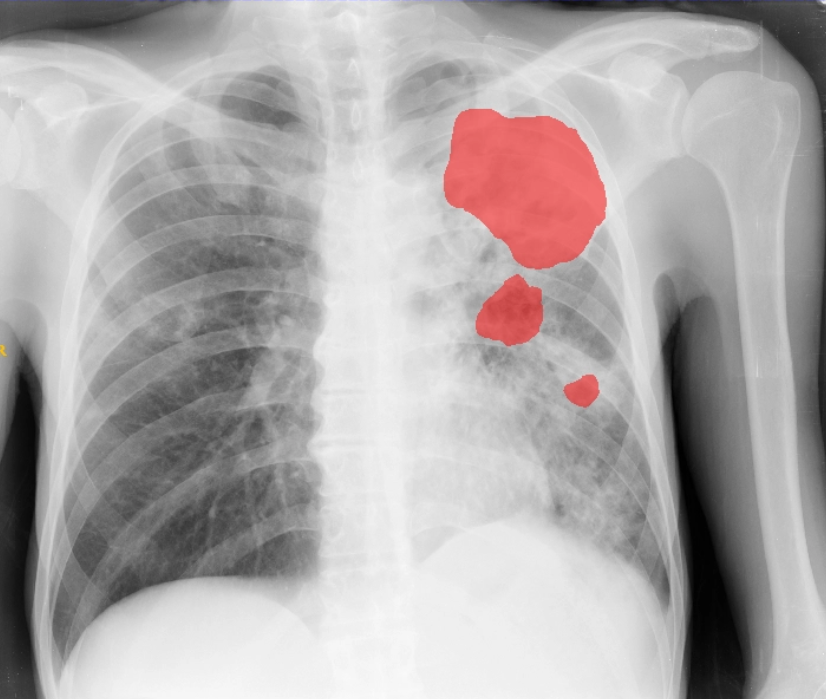
Why cavities matter — The presence of a thick-walled cavitary lesion in the upper lobes strongly favors active pulmonary tuberculosis, particularly post-primary (reactivation) disease. Such cavitation typically reflects caseous necrosis with subsequent drainage into the bronchial tree, creating an air-filled space that serves as a surrogate marker of high mycobacterial burden.
Since these cavitary foci frequently communicate with airways, patients with cavitary TB are more likely to exhibit smear- and culture-positive sputum, making the lesion a critical indicator of increased infectiousness and heightened transmission risk. While these radiological features are crucial indicators of TB severity, visual interpretation of CXRs is time-intensive and dependent on radiologist expertise, which may not always be available.
Deep learning–based detection and segmentation of cavitary lesions from CXR, targeting high-risk indicators of TB severity and infectiousness.
TB/Normal classification through multimodal fusion of CXR imaging and clinical metadata across multi-center international data.
External validation across 5 international sites — Korea, Mongolia, Peru, and the Philippines — ensuring clinical generalizability.
Build a model that determines whether a cavitary lesion is present on a frontal chest X-ray and segments the lesion when present..
Cavities are a key imaging marker of pulmonary tuberculosis severity and are strongly associated with bacterial burden, transmission risk, and treatment outcome. Automated cavity analysis can support more standardized and scalable TB assessment.
Participants must address two linked tasks:
Your method should be robust to variation in lesion appearance, size, location, and image quality.
For your final submission, please provide:
Detailed submission format specifications (file naming, directory structure, mask format) will be provided upon dataset release on May 22, 2026.
Build a model that classifies TB/Normal by integrating frontal chest X-ray imaging with clinical metadata.
Accurate classification of TB is fundamental to screening, treatment planning, and disease management, yet current practice relies heavily on subjective radiological interpretation. A standardized, quantitative measure of disease extent remains difficult to obtain manually and is prone to inter-observer variability. A multimodal AI framework that combines imaging features with clinical context — symptoms, demographics, bacterial load, medical history — can emulate the integrative reasoning of expert clinicians and improve diagnostic consistency across diverse healthcare settings.
Participants must develop a multimodal classification pipeline that takes two inputs simultaneously:
The model must accurately classify TB-positive and normal cases. Your method should be robust to missing or incomplete clinical variables and generalizable across diverse patient populations.
For your final submission, please provide:
Detailed submission format specifications and clinical metadata schema will be provided upon dataset release on May 22, 2026.
| Date | Event |
|---|---|
| Apr 10 – July 31, 2026 | Application for Participation |
| May 22, 2026 | Internal Dataset Release |
| May 22 – July 24, 2026 | Internal Competition Phase |
| July 31, 2026 | Announcement of Qualified Teams for External Competition |
| Aug 1 – Aug 20, 2026 | External Competition Phase |
| Aug 20 – Aug 31, 2026 | Organizer Verification and Docker Evaluation |
| September 1, 2026 | Final Ranking Announcement and MICCAI Invitation |
| September 27, 2026 | Challenge at MICCAI 2026 and Award Ceremony |
| November 1, 2026 | Paper Submission Deadline |
Participants will develop and validate their models using the internal dataset provided by the organizers. Based on the ranking achieved in this stage, selected teams will advance to the next stage.
Submission deadline: July 24, 2026, 23:59 (KST)
Selected teams will participate in the external competition for final evaluation. During this stage, each team may submit up to two times per week and must provide a Dockerfile. All submitted Dockerfiles will be executed directly by the organizers for final ranking.
A sample Dockerfile and submission template will be provided via the GitHub repository before the external competition begins.
Submission deadline: August 20, 2026, 09:00 AM (KST)
Important: Submissions with Dockerfiles that fail to execute successfully will be considered invalid.
Part of the Joint Thematic Day — Building Inclusive and Efficient AI Technologies for Medical Imaging in Africa and Other Resource-Constrained Settings
Chairs: Namkung Kim & Sengjoo Park
| Time | Session |
|---|---|
| 8:00 – 8:05 | Opening Remarks |
| 8:00 – 8:20 | (Keynote) Hoon Sang Lee (Right Foundation) — Bridging the Diagnostic Gap: Practical AI Deployment for TB in the Global South |
| 8:20 – 8:40 | Radiologists at the Forefront of Tuberculosis Eradication |
| 8:40 – 9:00 | TREAT-MMTB 2026 Challenge Overview |
| 9:00 – 10:00 | Top-Performing Teams Presentations |
| 10:00 – 10:30 | Coffee Break & Networking |
This session is part of the MICCAI 2026 Joint Thematic Day. Full agenda and venue details will be available on the MICCAI 2026 website. Schedule is subject to change.
Evaluation is based on Dice Similarity Coefficient (DSC) for segmentation quality and Accuracy for cavity presence detection.
Evaluation is based on F1-Score, balancing precision and recall to reflect clinical diagnostic requirements.
Submission format and file structure will be provided upon dataset release on May 22, 2026.
Docker base image specifications, submission template, and detailed guidelines will be provided via the GitHub repository before the external competition begins.
Use of additional publicly available datasets and pre-trained models is permitted. Participants may leverage any open-source datasets, foundation models, or pre-trained weights to improve their methods. All data sources and models used must be fully disclosed in the Method Description. Failure to disclose will result in disqualification.
Participants are free to use any computational resources during training. However, submitted inference pipelines must run within the organizers' evaluation environment. Models that exceed available GPU memory or fail to complete inference within the allotted time limit will be considered invalid. Specific hardware specifications and time constraints will be announced upon dataset release.
Top-ranking teams must submit full source code and trained model weights for verification. Any form of data leakage, test set probing, or result manipulation will result in immediate disqualification.
Teams or individuals may enter one or both tasks. Winning prizes in both tasks simultaneously is allowed. Each individual may belong to only one team per task. Participating in multiple teams within the same task is strictly prohibited. Team roster changes are permitted only once and must be finalized before the application deadline (July 31, 2026).
2nd Stage: Up to two submissions per week. Submissions with Dockerfiles that fail to execute will be considered invalid.
The organizers will provide a subset of annotated cases for training and validation. The source images originate from publicly available repositories, which participants may independently access. However, the annotations and labels created by the organizers are proprietary to this challenge and must not be redistributed or made publicly available at any time, including after the competition concludes. All participants are required to sign a data security agreement upon registration.
Participating teams may publish their results independently. Please cite the challenge paper when applicable.
Top three performing methods will be announced publicly on the challenge website. Selected teams will be invited to present their methodology during an oral presentation at the MICCAI 2026 Challenge Day.
These teams will be listed as co-authors in the final TREAT-MMTB journal manuscript. At a minimum, the top-performing teams selected to present at the workshop will be included. Additionally, submissions that exhibit notable scientific value may also be invited as co-authors in the journal publication, regardless of final ranking.
Winners are determined solely by each task's respective metrics; there is no combined overall ranking across tasks. To be eligible for any challenge awards, top-ranking teams are strictly required to publicly release their complete inference pipelines — including Dockerfile, preprocessing, postprocessing — and model weights.
Registration is free. You may register for one or both tasks.
Register via Google Form ↗Google Form — team info + data agreement
Download instructions + baseline code
Submit predictions
Docker + Method Description
Academic & Society
Radiology
Funding Support
Industry Partners

| What | When | Includes |
|---|---|---|
| - | - | - |
| - | - | - |
For questions or collaboration inquiries: [mi2rl.challenge@gmail.com]